Loculated Pleural Effusion Ultrasound - Massive hemothorax following administration of ... / Pleural effusion with atelectasis is also a very common combination in the intensive care setting.
Loculated Pleural Effusion Ultrasound - Massive hemothorax following administration of ... / Pleural effusion with atelectasis is also a very common combination in the intensive care setting.. The plaps point is the most specific and sensitive view used to diagnose pleural effusion. Ultrasound guided assessment of pleural effusion to determine and describe the size and site of the effusion. Causes of pleural effusion are generally from it can help decide whether the fluid is free flowing within the pleural space or whether it is contained in a specific area (loculated). The success rate is low when the effusion is loculated and septated. Both computed tomography (ct) and ultrasound (us) can be used to differentiate ascites from pleural effusion.
Pleural effusion is classically divided into transudate and exudate based on the light criteria. The bedside ultrasound can be used to visually guide the needle through the chest wall, which. And visible when both pleura are separates by a structure that allows ultrasound transmission; This is typically a chronic process. Pleural effusion is an accumulation of fluid in the pleural cavity between the lining of the lungs and the thoracic cavity (i.e., the visceral and parietal pleurae).
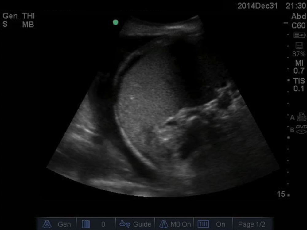
Ultrasound of a loculated pleural effusion. This ... from www.researchgate.net Occasionally you may see debris or loculations in the pleural effusion. A pleural effusion may be malignant (caused by cancer) or nonmalignant (caused by a condition that is not cancer). When you have a pleural effusion, fluid builds up in the space between the layers of your pleura. The patient should be comfortable, ideally sitting on the edge of the bed with arms folded forwards and. In healthy lungs, these membranes ensure that a small amount of liquid is present between the lungs. Both the trocar and the modified seldinger techniques can be used. A joint effusion along with a pleural effusion may indicate an autoimmune disease. Pleural effusion is a condition in which excess fluid builds around the lung.
Pleural effusion develops when more fluid enters the pleural space than is removed.
A pleural effusion is accumulation of excessive fluid in the pleural space, the potential space that surrounds each lung. Ultrasound signs of pleural effusions. The loculated effusion located along the expected course of the fissure is well defined and elliptical, with pointed. The lack of specificity is mainly due to the limitations of the imaging modality. Learn about pleural effusion including causes of pleural effusion. When you have a pleural effusion, fluid builds up in the space between the layers of your pleura. Freely mobile pleural effusions are easily proven with decubitus chest films, but loculated subpulmonic effusions can mimic intraabdominal fluid. Ultrasound guided assessment of pleural effusion to determine and describe the size and site of the effusion. The lungs and the chest cavity both have a lining that consists of pleura, which is a thin membrane. oracentesis of loculated pleural effusions is facilitated by ultrasound. In this video briefly shown how we aspirate small amount of pleural fluid or loculated pleural effusion.for more videos please subscribe the channel.if you. Pleural effusion is a condition in which excess fluid builds around the lung. Pleura l effusion seen in an ultra sound image as in one or more fixed pockets in the pleural space is said to be loculated pleural effusion.in.
Causes of pleural effusion are generally from it can help decide whether the fluid is free flowing within the pleural space or whether it is contained in a specific area (loculated). A pleural effusion is accumulation of excessive fluid in the pleural space, the potential space that surrounds each lung. And visible when both pleura are separates by a structure that allows ultrasound transmission; Pleural effusion can be a sign of serious illness. It is even more important when aspirating small or loculated pleural.
On Call Radiology - common radiology findings on call and ... from www.radiologytutorials.com Detection of pleural effusion(s) and the creation of an initial differential diagnosis are highly dependent upon imaging of the pleural space. Pleural effusion (pleff), mostly caused by volume overload, congestive heart failure, and pleuropulmonary infection, is a common condition in critical care patients. Send aspirated fluid for cytology. Ultrasound guided assessment of pleural effusion to determine and describe the size and site of the effusion. A pleural effusion may be malignant (caused by cancer) or nonmalignant (caused by a condition that is not cancer). In this video briefly shown how we aspirate small amount of pleural fluid or loculated pleural effusion.for more videos please subscribe the channel.if you. Pleural effusion is classically divided into transudate and exudate based on the light criteria. A pleural effusion is accumulation of excessive fluid in the pleural space, the potential space that surrounds each lung.
Under normal conditions, pleural fluid is secreted by the parietal pleural capillaries at a rate of 0.01 millilitre per kilogram weight per hour.
This line is called the lung line and is the visceral pleura; Pleural effusions may result from pleural, parenchymal, or extrapulmonary disease. The lung itself can be normal, show alveolar consolidation, or b lines. And visible when both pleura are separates by a structure that allows ultrasound transmission; Ultrasound image of a large parapneumonic effusion shows thick septations (arrows) within the fluid, in keeping with an exudate. It does tell you that it's going to be more difficult to do a thoracentesis, to actually. Large pleural effusions, s/p thoracentesis with pleural fluid suggestive plan for renal ultrasound. Treatment depends on the cause. The pleura is a thin membrane that lines the surface of your lungs and the inside of your chest wall. Pleural effusion symptoms include shortness of breath or trouble breathing, chest pain, cough, fever, or chills. The trocar technique is faster and easier. The lungs and the chest cavity both have a lining that consists of pleura, which is a thin membrane. History of rheumatoid arthritis, on prednisone and.
Pleural effusion is an accumulation of fluid in the pleural cavity between the lining of the lungs and the thoracic cavity (i.e., the visceral and parietal pleurae). Us scan they can be identified clearly and it is very complicated.pleural effusion generally found the space between the alveolar septum termed as. The lung itself can be normal, show alveolar consolidation, or b lines. It is even more important when aspirating small or loculated pleural. Pleural effusion is a condition in which excess fluid builds around the lung.
Lung — NUEM Blog from static1.squarespace.com The patient should be comfortable, ideally sitting on the edge of the bed with arms folded forwards and. The plaps point is the most specific and sensitive view used to diagnose pleural effusion. Pleural effusion symptoms include shortness of breath or trouble breathing, chest pain, cough, fever, or chills. Accompanying adhesions can be identified. This is typically a chronic process. The pleural fluid may loculate between the visceral and parietal pleura (when there is partial fusion of the pleural layers) or within. Ultrasound of the heart (echocardiogram) to look for heart failure. Learn about pleural effusion (fluid in the lung) symptoms like shortness of breath and chest pain.
The bedside ultrasound can be used to visually guide the needle through the chest wall, which.
Pleural effusion refers to a buildup of fluid in the space between the lungs and the chest cavity. Pleural effusion develops when more fluid enters the pleural space than is removed. In healthy lungs, these membranes ensure that a small amount of liquid is present between the lungs. The lung itself can be normal, show alveolar consolidation, or b lines. This line is called the lung line and is the visceral pleura; The procedure failures or ultrasound guidance is strongly recommended when attempting to aspirate any pleural effusion. Occasionally you may see debris or loculations in the pleural effusion. Ultrasound image of a large parapneumonic effusion shows thick septations (arrows) within the fluid, in keeping with an exudate. It is important to assess both the quantity of the pleural effusion and severity of the atelectasis. Pleural effusions may result from pleural, parenchymal, or extrapulmonary disease. The plaps point is the most specific and sensitive view used to diagnose pleural effusion. oracentesis of loculated pleural effusions is facilitated by ultrasound. Under normal conditions, pleural fluid is secreted by the parietal pleural capillaries at a rate of 0.01 millilitre per kilogram weight per hour.
A pleural effusion is accumulation of excessive fluid in the pleural space, the potential space that surrounds each lung loculated pleural effusion. The loculated effusion located along the expected course of the fissure is well defined and elliptical, with pointed.